Radiology Blog cafe

xray, CT and MRI radiology, CT and MR image fusion with special augmented reality techniques like volume rendering,3D SSD using dedicated work stations playing with volume data sets offering surgeon friendly images with structured reporting is the modern Radiology.radiology blog cafe will high light clinical cases with augmented reality impacts with interactive educative blogs.

Sunday, August 06, 2006

virtual ventriculoscopy


virtual ventriculoscopy : 3d volume rendered virtual fly through navigation image from 3D FSE data set of a 3rd ventricular germ cell tumor seen on T2 sagittal and FLAIR axial at the posterior 3rd ventricle with obstructive hydrocephalous.Note the MR spectroscopy image showing very high choline creatine ,high lipid lactate with very Low NAA.

conjoint twins


volume rendered CT of conjoint twins done on SIEMENS sensation sensation 16 and reconstructed on WIZARD showing the abnormally fused skeleton and anomalies in these conjoint twins sharing organs.

3D CT and MRI image fusion


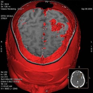
3D volume rendered CT+MR fused image showing the AVM in left posterior superior parietal convexity.


Original CT angio volume rendered image shows AVM nidus with arterial feeders and draining veins


augmented reality


diffusion tensor imaging with fiber tracking showing thalamic radiations http://www.ismrm.org